Shoulder Injuries
Rotator cuff muscles
Supraspinatus (Abductor) – abduction 20 – 40 in extreme internal rotation with palms outwards
Subscapularis (internal rotator) – extending hand away from back.
Infraspinatus & Teres minor – external rotation
A combination of 4 tests can be used to assess the strength of the rotator cuff: –
- The empty-can test evaluates the supraspinatus.
- The patient raises both arms slightly forward from the coronal plane of the trunk with thumbs pointing to the floor.
- The examiner applies pressure to the top of the arms, which the patient attempts to resist.
- Weakness indicates a supraspinatus tear.
- The external rotation test isolates the infraspinatus.
- With the arm at his or her side and the elbow flexed to 90°, the patient attempts to externally rotate against resistance supplied by the examiner.
- Infraspinatus tears result in pain and weakness.
- The lift-off test – for subscapularis
- evaluates the patient’s ability to lift the hand away from the small of the back as the examiner applies resistance.
- Weakness suggests a subscapularis tear.
- The belly-press test – Subscapularis
- the patient presses the hand against the umbilicus with the elbow forward from the trunk.
- The examiner applies resistance by placing a hand between the patient’s hand and abdomen.
- Inability to maintain elbow anterior to the coronal plane of the trunk suggests a subscapularis tear.
Subacromial impingement (SAI)
It is due to impingement of the supraspinatus beneath the acromion.
U/S determines if a tear is present.
Treatment include Rest, Analgesic (NSAID) and physiotherapy. If failed – Arthroscopic surgery
Clinical examination: –
- Neer’s Test: “passive painful arc manoeuvre” The examiner should stabilize the patient’s scapula with one hand, while passively flexing the arm while it is internally rotated (from 60 to 120°). If the patient reports pain in this position, then the result of the test is considered to be positive.
- Jobe Test: – Pain without weakness suggests tendinopathy; pain with weakness is consistent with tendon tear.
Acromioclavicular (AC) joint injury
Common injuries follow fall onto the shoulder or violent sudden movements of the upper limb.
Treatment: – analgesia, support in a broad arm sling, and arrange follow-up after 3 weeks.
AC joint injuries are classified:
- Type I: minimal separation. Acromioclavicular (AC) Ligament Sprain, Coracoclavicular (CC) Ligament Intact and Joint Capsule Intact.
- Type II: obvious subluxation, AC Ligament Rupture, CC Ligament Sprain and Joint Capsule Rupture.
- Type III: complete dislocation of AC joint. AC /CC Ligaments Rupture and Joint Capsule Rupture.
- Type IV: The clavicle is displaced posteriorly. AC /CC Ligaments Rupture and Joint Capsule Rupture.
- Type V: displacement is > 100 %. AC /CC Ligaments Rupture and Joint Capsule Rupture.
- Type VI: The Clavicle is displaced inferiorly. AC /CC Ligaments Rupture and Joint Capsule Rupture.

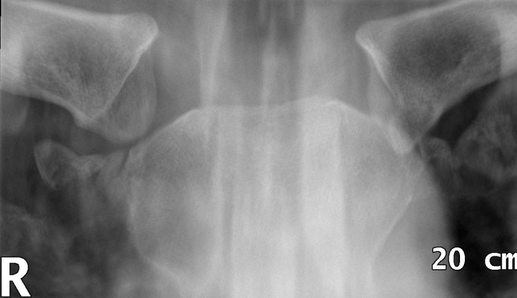
Scapular fracture
Direct falls on to the back such as being thrown from a horse. The injury may not be noticed if accompanied by other, more severe injuries.
Diagnosis may require a skyline view
Treatment involves pain control, immobilizing the affected area and subsequently physiotherapy.
Winging of the scapula
Diagnosed clinically when patient presenting with shoulder pain and weakness.
Caused by serratus anterior muscle dysfunction due to muscle injury or injury to the nerve supply – The long thoracic nerve.
The specific examination test is serratus wall test
Treatment is mainly by physiotherapy and range of motion exercises, if failed after 6 – 12 months surgical treatment is needed.
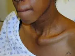
Sternoclavicular disruption
Direct force to the front of this joint can cause a posterior dislocation, one of the few upper limb injuries that can cause an immediate threat to life.
It can give rise to cough, hoarseness and even pneumothorax or tracheal compression.
It can cause ipsilateral arm venous congestion due to compression of the internal jugular vein.
An attempt can be made for closed reduction. Traction is applied to the arm then try to grasp the clavicle through the skin & pull it forwards – hopefully resulting in a pop as reduction occurs.
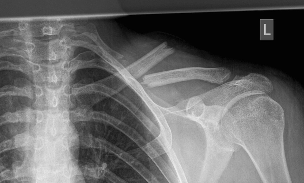
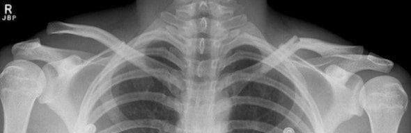
Clavicle fracture
The most common bones to be fractured, most often in the middle third.
Children are particularly prone to the fracture, and new-borns may present with a clavicle fracture following a difficult delivery.
Treatment of the uncomplicated fracture is generally to provide a broad arm sling, analgesia and fracture clinic for monitoring progress with X-rays.
Surgery is indicated when one or more of the following conditions presents:
- Comminution with separation
- Significant shortening of the clavicle
- Skin penetration
- Associated neurological or vascular injury.
- Non-Union after 3 – 6 months
Anterior Shoulder dislocations
Mechanism of injury: Forced external rotation/abduction of the shoulder.
Examination:
- “Cows bottom appearance” From the rear.
- Step-off deformity at the acromion with palpable gap below the acromion.
- Humeral head palpable antero-inferiorly to the glenoid.
- Arm is slightly abducted and externally rotated.
X-ray shows: –
- Loss of congruity between humeral head and the glenoid.
- inferomedial displacement of the humeral head on AP shoulder X-ray.
- “Hill-Sachs deformity” seen more in the axial view
- “Bankart lesion” due to tear to lower part of the glenoid labrum


Complications:
- Brachial plexus injury or axillary nerve injury
- Axillary artery or vein compression
- Rotator cuff tears /Hill-Sachs lesion & Bankart lesion
- Shoulder instability and recurrence of dislocation
Treatment:
- Check distal pulses and sensation over the lateral aspect of the shoulder before & after reduction.
- Reduce under Entonox® alone or sedation/analgesia with full monitoring.
- Immobilize in broad arm sling or collar & cuff for 3 weeks. Check X-ray after reduction.
- Provide analgesia and fracture clinic follow-up.
Methods of reduction: –
- The Spaso manoeuvre: – Patient lies supine, the upper limb is held externally rotated by the body, in gentle traction, then gradually flexed through, if necessary, 180 degrees.
- External rotation method: – With the patient’s arm adducted and the elbow flexed, the forearm is slowly and gently externally rotated (without force or traction). If pain or spasm is felt, the physician stops and allows the patient to relax.
- Modified Milch method: Slowly abduct the straight arm to 110⁰. With the elbow extended, apply gentle steady traction to the arm, while an assistant controls movement of the humeral head back into the glenoid.
- Scapular manipulation methods: With the patient lying prone, ‘manipulate’ the scapula onto the glenoid by pushing the inferior tip of the scapula medially and the superior part laterally.
- Stimson’s technique: A more traditional method with the patient prone. Apply a weight strapped to the forearm/wrist of the affected side as it hangs down and await reduction.
Posterior Shoulder dislocations
Mechanism of injury: They occur with forced internal rotation & adduction of the shoulder. They are associated with seizures and electrocution.
On examination: – The patient loses the ability to externally rotate.
X-ray shows: –
- absence of external rotation on images in a standard shoulder series is a clue.
- The light bulb sign seen on an AP view of the shoulder.
- loss of normal half-moon overlap sign, in which the glenoid fossa appears vacant due to the lateral displacement of the humeral head.

Associated injuries: –
- Tuberosity and surgical neck fractures of the humerus
- Reverse Hill-Sachs lesions / Injuries to the Labrum
- Rotator cuff injuries
Treatment:
- Check neurovascular status.
- Manipulate under sedation by applying traction & external rotation to the upper limb at 90⁰. If difficult, refer for reduction under GA.
| Adhesive capsulitis (frozen shoulder) | Calcific tendonitis |
| This usually results from trauma which in turn leads to capsular contraction. Clinical features: – there is globally reduced ROM, with enhanced scapulo-thoracic movements as the patient can only abduct in this way. Clinical examination: The hands-on-hip test (Sloans test) is helpful. It should be possible to move the elbows more anterior to the fixed wrists. Treatment: Steroid & Judicious physiotherapy can limit capsular tightness, but manipulation under anaesthetic (MUA) produces good results with many patients gaining an almost full ROM in 2 – 3 months | # This typically occurs in young to middle aged adults with no previous problem. # There is a sudden onset of excruciating pain, typically preventing sleep. This is due to leakage of calcium deposits from the supraspinatus tendon into the joint space. # In most cases, clinical symptoms will resolve spontaneously in 7 – 10 days. |